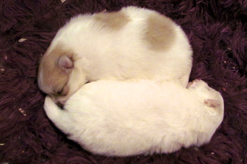
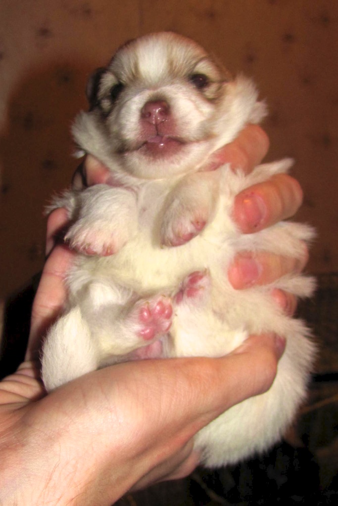
The puppies opened their eyes recently. They don’t look like puppies, in my opinion; more like smug little bears. The brown and white one yawns a lot. They are of course still suckling, though Bebe is much less protective of them and checks in on them only occasionally. That said, if she spots any of the other poms going into the bedroom, there’s always pomeranian drama, lol. Puppy pictures are below ^-^
My plants are doing well. I finally decided to cut back the belladonna, which almost immediately grew new leaves from the 1/2″ stems I left. I’ve been wanting to start some Black Pearl Peppers (the leaves are black instead of green) and Easter Egg Radishes, the latter of which may help me with one of my graduate projects. I transplanted the belladonna outside when it was relatively warm the other day. I’m hoping the roots may survive the rest of the Maine winter, especially since the species is native to Europe. I also have some blue water lilies growing and although their stems are about a foot long now, their growth, much like that of the Colchicaceae plants, has virtually halted, though they remain healthy. I have them under a grow light, but I’m not sure it’s helping.
Two of my three orchids are shooting, an odd time considering the light is at a minimum right now. One of them, an Encyclia orchid I got from R.F. Orchids in Florida nearly two years ago, will bloom for the first time in its life. The flowers supposedly smell of chocolate, so I’m excited! I will post pictures when they bloom.
Among my Christmas presents were two microscopes – a dissecting scope and an objective microscope with 2,000X power. It’s my fourth objective microscope, but this one includes an attachment for dark-field microscopy, which is great for certain microorganisms with transparent bodies. I had to cut some infected leaf tissue from one of my Phalaenopsis orchids the other day and I was able to view the stomata, chloroplasts, nucleus, and nucleolus very clearly even though I don’t have any stains yet. Just by adjusting the diaphragm, I was able to observe changes in the cytoplasmic streaming of the chloroplasts.
Unfortunately UMaine doesn’t offer any courses in plant histology, so I’m on my own when it comes to using my microtome and figuring out what stains I need to order. There are a lot of good resources online, though. I’ll post pictures when I get some good ones. I also ordered a camera mount while I was in California, so now I can take time-lapse videos. I’m hoping to (very carefully) slice open a dicot plant seed in order to photograph the embryonic developmental stages (embryogenesis), which are very distinctive. This Summer I’ll likely take some time to document the cleavage stages of amphibian eggs and their embryonic development as well. In the meantime there’s limitless things to look at for fun, like my red and white blood cells, cheek cells, hair, and microorganisms, many species of which I can find in the standing water around my orchids. Sometimes I’m lucky enough to come across Daphnia, my favorite. I bet they will show up well under the dark field.